

S-Gal (3,4-Cyclohexenoesculetin-β-D-galactopyranoside, CAS 182805-65-8), commercially known as S-Gal®, represents a superior chromogenic substrate to X-Gal for β-galactosidase (β-Gal, EC 3.2.1.23) detection, hydrolyzing the β-glycosidic bond to release 3,4-cyclohexenoesculetin that chelates ferric ions (Fe³⁺) forming an intense black insoluble precipitate with strong T₂* MRI contrast enhancement, enabling both histological visualization and noninvasive molecular imaging. With molecular formula C19H22O9 (MW 394.38 g/mol), this white crystalline solid features β-D-galactopyranose linked at C1-O to the enol form of cyclohexenoesculetin—a coumarin derivative with vicinal hydroxy groups at positions 3,4—exhibiting rapid cleavage by E. coli lacZ (α-complementation), mammalian lysosomal β-galactosidases, and viral β-Gals (kcat/Km >10⁴ M⁻¹s⁻¹), producing the catechol-like aglycone that quantitatively binds Fe³⁺ (1:2 stoichiometry) to generate superparamagnetic nanoscale precipitates detectable by eye, absorbance (λmax 590 nm), and MRI (ΔR₂* >100 s⁻¹mM⁻¹). Unlike X-Gel’s diffusion-prone indigo, S-Gal’s nondiffusible black product localizes precisely to enzyme sources, supporting high-density colony screening (>10⁴ CFU/plate) with 2-4x faster color development (visible at 4h vs. 16h), while Fe³⁺ complexation enables multimodal readout (histology/MRI/optical) for in vivo reporter gene imaging in lacZ-transfected tumor xenografts. Used at 100-200 μg/mL with 0.5-2 mM ferric ammonium citrate (FAC), it revolutionizes molecular imaging by bridging traditional histology with clinical MRI, with applications in gene therapy monitoring, oncolytic virotherapy tracking, and real-time β-Gal pharmacodynamics without ionizing radiation.
Appearance
Source
Molecular Weight and Structure
Sugar Specificity
Biological Activity
Purity and Microbial Contamination
Identity and Quality Control
Shelf Life and Storage
Applications
Key Characteristics
Citation Links
| Name | CNP-α-L-Fuc |
| Synonyms | 2-Chloro-4-nitrophenyl-α-L-fucopyranoside |
| CAS Number | 157843-41-9 |
| Molecular Formula | C12H14ClNO7 |
| Molecular Weight | 319.70 g/mol |
| Specifications & Purity | 98% (Vacuum foil packaging) |
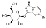
| Name | Salmon-Gal |
| Synonyms | 6-Chloro-3-indolyl-β-D-galactopyranoside |
| CAS Number | 138182-21-5 |
| Molecular Formula | C14H16ClNO6 |
| Molecular Weight | 329.73 g/mol |
| Specifications & Purity | ≥95% (Vacuum foil packaging) |
| Name | CNP-GlcNAc |
| Synonyms | 2-Chloro-4-nitrophenyl β-D-N-acetylglucosaminide |
| CAS Number | 103614-82-0 |
| Molecular Formula | C14H17ClN2O8 |
| Molecular Weight | 376.75 g/mol |
| Specifications & Purity | 98% (Vacuum foil packaging) |
| Name | Magenta-GlcA Sodium Salt |
| Synonyms | 5-Bromo-6-chloro-3-indolyl-β-D-glucuronide sodium salt |
| CAS Number | 216971-54-9 |
| Molecular Formula | C14H12BrClNNaO7 |
| Molecular Weight | 444.59 g/mol |
| Specifications & Purity | 98% (Vacuum foil packaging) |
Reviews
There are no reviews yet.