")
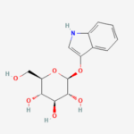
Y-α-D-Glc (3-Indolyl-α-D-glucopyranoside, CAS 914912-11-1) serves as a chromogenic substrate specifically designed for the detection of α-glucosidase (EC 3.2.1.20) enzymes, where hydrolysis of the α-glycosidic linkage releases 3-indoxyl that undergoes rapid spontaneous oxidation and dimerization to form an insoluble indigo-blue precipitate, enabling straightforward visual screening of enzyme-positive bacterial colonies, fungal cultures, or recombinant plaques without requiring specialized equipment. With molecular formula C14H17NO6 and molecular weight of 295.29 g/mol, this compound consists of an α-D-glucopyranosyl moiety O-linked at the anomeric C1 position to the 3-hydroxy group of unsubstituted 1H-indole, distinguishing it from its β-anomer counterpart (Y-β-D-Glc) by its strict specificity for α-glucosidases from sources such as Saccharomyces cerevisiae maltase, human intestinal sucrase-isomaltase, and Bacillus stearothermophilus α-glucosidase, while exhibiting minimal cross-reactivity with β-glucosidases due to the axial anomeric configuration. The unsubstituted indole ring confers faster oxidation kinetics compared to halogenated derivatives like X-α-Glc, resulting in intensified blue color development within 12-24 hours at concentrations of 100-300 μg/mL in agar media, though its higher propensity for spontaneous hydrolysis necessitates preparation of fresh solutions to minimize background staining. This substrate finds primary utility in food microbiology for identifying α-glucosidase-positive probiotics and pathogens involved in starch/sucrose fermentation, as well as in industrial biotechnology for screening engineered GH13 family glycoside hydrolases in bioethanol production and functional characterization of microbial α-glucosidases in complex carbohydrate degradation pathways.
Appearance
White to pale beige crystalline powder, hygroscopic in nature. Hydrolysis produces characteristic insoluble blue-indigo precipitate visible under ambient light.
Source
Synthesized via Koenigs-Knorr glycosylation employing protected α-D-glucopyranosyl bromide with indol-3-ol under silver oxide catalysis, followed by deprotection. Available from specialty fine chemical suppliers focused on glycobiology reagents.
Molecular Weight and Structure
Molecular formula C14H17NO6; exact mass 295.29 g/mol. Systematic name: (2R,3S,4R,5R,6R)-2-(1H-indol-3-yloxy)-6-(hydroxymethyl)tetrahydro-2H-pyran-3,4,5-triol, featuring α1-O-glycosidic linkage confirmed by ¹H-NMR anomeric proton doublet at δ ~5.3 ppm (J = 3.5 Hz).
Sugar Specificity
Highly selective for α-D-glucopyranosidases (GH13 family), with >100-fold preference over β-glucosidases; negligible reactivity with galactosidases or fucosidases.
Biological Activity
Chromogenic reporter substrate with Km values typically 0.3-1.0 mM for microbial α-glucosidases; liberates indoxyl for oxidative dimerization to indigo (λmax ~600 nm), enabling colony visualization without IPTG induction.
Purity and Microbial Contamination
≥98% purity by HPLC; synthesized as research chemical with no microbial contamination, though hygroscopic nature requires desiccated handling.
Identity and Quality Control
Confirmed by ¹H-NMR (indole NH ~10.8 ppm, α-anomeric H ~5.3 ppm), HRMS m/z 296.1 [M+H]⁺, and specific rotation [α]D +120° (c=1, DMF); single TLC spot (Rf 0.4, EtOAc:MeOH 9:1).
Shelf Life and Storage
Stable for 2+ years when stored desiccated at -20°C; DMSO or DMF stocks (50 mM) viable for 6 months at -80°C, protect from moisture and light exposure.
Application
Screening α-glucosidase-positive probiotics and pathogens in dairy/food microbiology; functional assays for GH13 enzymes in starch bioethanol production; orthogonal screening to β-glucoside substrates in glycosidase libraries.
Key Characteristics
Rapid blue color development (12-24h) versus halogenated analogs; cost-effective unsubstituted indole scaffold; water-miscible for easy media incorporation; α-specific orthogonal to common β-substrates.
Citation Links
https://www.chemicalbook.com/ChemicalProductProperty_EN_CB34656002.htm
https://amp.chemicalbook.com/ChemicalProductProperty_EN_CB34656002.htm
https://www.chemicalbook.com/ProductChemicalPropertiesCB34656002_EN.htm
https://www.sigmaaldrich.com (indolyl glucoside catalog)
https://www.tcichemicals.com (α-glucosidase substrates)
https://www.biosynth.com (chromogenic glycosides)
https://www.medchemexpress.com (enzyme substrates)
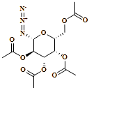
| Cas No : | 13992-26-2 |
| Mol Formula & Weight : | C14H19N3O9, 373.32 |
| IUPAC Name : | [(2R,3S,4S,5R,6R)-3,4,5-triacetyloxy-6-azidooxan-2-yl]methyl acetate |
| Purity : | >99% (HPLC) |
| Synonyms : | 13992-26-2, 1-Azido-1-deoxy-beta-D-galactopyranoside tetraacetate, 2,3,4,6-tetra-O-acetyl-beta-D-galactopyranosyl azide, [(2R,3S,4S,5R,6R)-3,4,5-triacetyloxy-6-azidooxan-2-yl]methyl acetate |
10 in stock
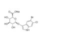
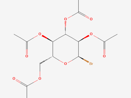
| Cas No : | 572-09-8 |
| Mol Formula & Weight : | C14H19BrO9, 411.2 |
| IUPAC Name : | [(2R,3R,4S,5R,6R)-3,4,5-triacetyloxy-6-bromooxan-2-yl]methyl acetate |
| Purity : | >99% (HPLC) |
| Synonyms : | 572-09-8, 2,3,4,6-Tetra-O-acetyl-alpha-D-glucopyranosyl bromide, Acetobromglucose, acetobromoglucose |
10 in stock
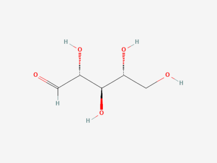
| Cas No : | 50-69-1 |
| Mol Formula & Weight : | C5H10O5, 150.13 |
| IUPAC Name : | (2R,3R,4R)-2,3,4,5-tetrahydroxypentanal |
| Purity : | >99% (HPLC) |
| Synonyms : | 50-69-1, (2R,3R,4R)-2,3,4,5-tetrahydroxypentanal, aldehydo-D-ribose, Ribose, D- |
10 in stock
| Cas No : | 4163-65-9 |
| Mol Formula & Weight : | C16H22O11, 390.34 |
| IUPAC Name : | [(2R,3R,4S,5S,6R)-3,4,5,6-tetraacetyloxyoxan-2-yl]methyl acetate |
| Purity : | >99% (HPLC) |
| Synonyms : | 4163-65-9, a-D-Mannose pentaacetate, 1,2,3,4,6-Penta-O-acetyl-alpha-D-mannopyranose, alpha-d-Mannose pentaacetate |
10 in stock


Reviews
There are no reviews yet.