
| Appearance | White amorphous powder, lyophilized | |
|---|---|---|
| Activity | GradeⅢ 150 U/mg-solid or more | |
| Contaminants | Phosphoglucose isomerase | ≤1.0×10⁻¹ % |
| 6-Phosphogluconate dehydrogenase | ≤1.0×10⁻² % | |
| Glucose-6-phosphate dehydrogenase | ≤1.0×10⁻² % | |
| Myokinase | ≤1.0×10⁻² % | |
| Glutathione reductase | ≤5.0×10⁻¹% | |
| Stability | Stable at −20 ℃ for at least one year(Fig.1) |
|---|---|
| Molecular weight | approx. 82,000 (by gel filtration) |
| Isoelectric point | 4.1±0.1 |
| Michaelis constants | 2.3×10⁻⁴ M (D-Glucose), 7.7×10⁻⁵ M (ATP) |
| Inhibitors | Metal ions, p-chloromercuribenzoate, iodoacetamide, SDS, etc |
| Optimum pH | 8.0−9.0(Fig.2) |
| Optimum temperature | 50 ℃(Fig.3) |
| pH Stability | pH 4.0−9.0 (25 ℃, 20 hr)(Fig.4) |
| Thermal stability | below 45 ℃ (pH 7.0, 30 min)(Fig.5) |
| Substrate specificity | (Table 1) |
| Effect of various chemicals | (Table 2) |
The enzyme is useful for enzymatic determination of glucose, adenosine-5′-triphosphate (ATP) and creatine phosphokinase in combination with glucose-6-phosphate dehydrogenase (=G-6-PDH, G6D311, G6D-321).
![]()
![]()
The formation of NADH is measured at 340 nm by spectrophotometry.
One unit causes the formation of one micromole of NADH per minute under the conditions detailed below.
| A. Tris-HCl buffer, pH 8.0 | 50 mM, containing 13.3 mM MgCl2 | |
|---|---|---|
| B. Glucose solution | 0.67 M in Tris-HCl buffer solution (A) (The solution should be keep at room temperature for at least 1 hour before use) | |
| C. ATP solution | 16.5 mM in Tris-HCl buffer solution (A) (Should be freshly prepared) | |
| D. NAD+ solution | 6.8 mM in Tris-HCl buffer solution (A) (Should be freshly prepared) | |
| E. G-6-PDH solution | 300 U/mL (Dilute with Tris-HCl buffer solution (A) and store on ice) | |
| F. Enzyme diluent | Tris-HCl buffer solution (A) containing 0.1 % of bovine serum albumin | |
1.Prepare the following reaction mixture in a cuvette (d= 1.0cm) and equilibrate at 30℃ for approximately 5 minutes.
| 2.30 mL | Tris-HCl buffer solution | (A) |
| 0.50 mL | Glucose solution | (B) |
| 0.10 mL | ATP solution | (C) |
| 0.10 mL | NAD+ solution | (D) |
| 0.01 mL | G-6-PDH solution | (E) |
| Concentration in assay mixture | |
|---|---|
| Tris-HCl buffer | 50 mM |
| Glucose | 0.11 M |
| ATP | 0.53 mM |
| NAD+ | 0.22 mM |
| MgCl2 | 13 mM |
| BSA | 3.2 μg/mL |
| G-6-PDH | approx.1.0 U/mL |
2.Add 0.1ml of the enzyme solution* and mix by gentle inversion.
3.Record the increase of optical density at 340 nm against water for 4 to 5 minutes with a spectrophotometer thermostated at 30 ℃ and calculate the ΔOD per minute from the initial portion of the curve (ΔOD test).
At the same time, measure the blank rate (ΔOD blank) by the same method as the test except that the enzyme diluent (F) is added instead of the enzyme solution.
*Dissolve the enzyme preparation on ice-cold enzyme diluent (F) and dilute to 0.1−0.3 U/mL with the same buffer, immediately before the assay.
Activity can be calculated by using the following formula :
Volume activity (U/ml) =
ΔOD/min (OD test−OD blank)×Vt×df
6.22×1.0×Vs
= ΔOD/min×5.0×df
Weight activity (U/mg) = (U/ml)×1/C
| Vt | : Total volume (3.11 mL) |
| Vs | : Sample volume (0.1 mL) |
| 1.0 | : Light path length (cm) |
| 6.22 | : Millimolar extinction coefficient of NADH (cm2/micromole) |
| df | : Dilution factor |
| C | : Enzyme concentration in dissolution (c mg/mL) |
[Pyruvate kinase-Lactate dehydrogenase system with 0.1 M Tris-HCl buffer, pH 7.5]
| Substrate(100mM) | Relative activity(%) |
|---|---|
| D-Glucose | 100 |
| D-Fructose | 140 |
| D-Mannose | 52 |
| 2-Deoxy-D-glucose | 91 |
| Substrate(100mM) | Relative activity(%) |
|---|---|
| D-Galactose | 0 |
| D-Xylose | 2 |
| D-Glucosamine | 58 |
[The enzyme dissolved in 50mM K-phosphate buffer, pH 6.5 (5 U/mL) contg. 0.1 % bovine serum albumin was incubated with each chemical at 30 ℃ for 1hr.]
| Chemical | Concn.(mM) | Residual activity(%) |
|---|---|---|
| None | – | 100 |
| Metal salt | ||
| AgNO3 | 2.0 | 0 |
| BaCl2 | 2.0 | 99 |
| CaCl2 | 2.0 | 98 |
| CdCl2 | 2.0 | 85 |
| CoCl2 | 2.0 | 85 |
| CuSO4 | 2.0 | 25 |
| FeCl3 | 2.0 | 28 |
| FeSO4 | 2.0 | 80 |
| HgCl2 | 2.0 | 0 |
| MgCl2 | 2.0 | 98 |
| MnCl2 | 2.0 | 100 |
| NiCl2 | 2.0 | 100 |
| Pb(OAc)2 | 2.0 | 98 |
| Zn(OAc)2 | 2.0 | 98 |
| ZnSO4 | 2.0 | 99 |
| NaF | 20.0 | 101 |
| NaN3 | 20.0 | 102 |
| Chemical | Concn.(mM) | Residual activity(%) |
|---|---|---|
| PCMB | 2.0 | 0 |
| MIA | 2.0 | 80 |
| IAA | 2.0 | 7 |
| EDTA | 5.0 | 103 |
| (NH4)2SO4 | 20.0 | 104 |
| Borate | 20.0 | 102 |
| o-Phenanthroline | 2.0 | 101 |
| α,α′-Dipyridyl | 2.0 | 102 |
| Urea | 2.0 | 104 |
| Guanidine | 2.0 | 103 |
| Hydroxylamine | 2.0 | 104 |
| Na-cholate | 1.0 % | 102 |
| Triton X-100 | 1.0 % | 105 |
| Brij 35 | 1.0 % | 0 |
| SDS | 0.1 % | 25 |
| Tween 20 | 0.1 % | 101 |
| Span 20 | 0.1 % | 106 |
| DAC | 0.1 % | 101 |
Ac, CH3CO; NEM, N-Ethylmaleimide; PCMB, p-Chloromercuribenzoate; MIA, Monoiodoacetate; EDTA, Ethylenediaminetetraacetate; IAA, Iodoacetamide; SDS, Sodium dodecyl sulfate; DAC, Dimethylbenzylalkylammonium chloride.
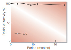
Fig.1. Stability (Powder form)
(kept under dry conditions)
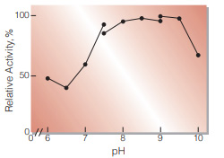
Fig.2. pH-Activity
30 ℃ in the 50 mM buffer solution: pH 6.2-7.5, PIPES-NaOH: pH 7.5-9.0, Tris-HCI: pH 9.0-10.0, Glycine-NaOH
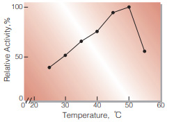
Fig.3. Temperature activity
(in 50 mM Tris-HCI buffer,pH 8.0)
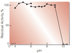
Fig.4. pH-Stability
25 ℃, 20 hr-treatment in the 0.1 M buffer solution: pH 4.0-8.0,Acetate-NaOH;pH 6.0-8.0, K-phosphate; pH 7.5-9.0,Tris-HCl;pH 9.0-10.5, Glycine-NaOH enzyme concn.: ca.10 U/mL
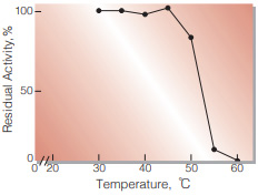
Fig.5. Thermal stability
30 min-treatment with 50 mM K-phosphate buffer, pH 7.0, containing 0.1% bovine serum albumin enzyme concn.: ca.5 U/mL
| Size | 1 MG, 10 MG, 5 MG |
|---|
| Name | α1,2-fucosyltransferase; α1,2FucT |
| Product Code | EN01013 |
| E.C. | 2.4.1.69 |
| Product Description | E. coli recombinant α1,2-fucosyltransferase from Helicobacter mustelae |
| Unit Definition | One unit is defined as the amount of enzyme that catalyzes the formation of 1μmol Fucα1,2Lac from GDP-Fuc and lactose per minute at 37 °C. |
10 in stock
| Name | Sialic acid aldolase (NPL) |
| Catalog No. | EN01027 |
| E.C. | 4.1.3.3 |
10 in stock
| Name | GlcNAc-1-P uridyltransferase (GlmU) |
| Catalog No. | EN01012 |
| E.C. | 2.3.1.157 |
10 in stock
| Name | Galactokinase; BiGalK |
| Product Code | EN01018 |
| E.C. | 2.7.1.6 |
| Product Description | E. coli recombinant galactokinase from Bifidobacterium infantis |
| Unit Definition | One unit is defined as the amount of enzyme that catalyzes the formation of 1 μmol of Gal-1-P from galactose and ATP per minute at 37 °C. |
10 in stock


Reviews
There are no reviews yet.