About this product
Introduction
Recent technological advances by have enabled the largest commercially available glycosylation antibody array to date. With the Human Glycosylation Array 8000, researchers can now efficiently obtain glycosylation profiles in their samples. The glycosylation profiles of 8000 human proteins can be simultaneously detected, including, but not limited to, cytokines, chemokines, adipokines, growth factors, angiogenic factors, proteases, soluble receptors, soluble adhesion molecules in cell culture supernatants, serum, plasma and other samples types.
Capture antibodies are printed onto glass slides and the glycans on these capture antibodies are removed. The glass slide arrays come pre-blocked and are ready to be incubated with samples. After incubation with samples and washing to remove unbound proteins, five unique biotin-labeled lectins are incubated with the array to allow binding to the glycans from captured target proteins. Streptavidin-conjugated fluorescent dye (Cy3 equivalent) is then added to the array. Finally, the glass slide is dried and laser fluorescence scanning is used to visualize the signals. These signals are then compared to the array map to identify glycosylated proteins present in the samples.
Product Features
High density screening
High sensitivity and specificity
Low sample volume (20-100 µl per array)
Large dynamic range of detection
Compatible with most sample types
Accurate and reproducible
Target Names
8,000 Target Protein Names and Identifiers [Excel Download]
Biotin-Labeled Lectin Mixture
Lectin Name
Sugar specificity
1
Concanavalin A
αMan, αGlc
2
Dolichos Biflorus Agglutinin
αGalNAc
3
Peanut Agglutinin
Galβ3GalNAc
4
Ulex Europaeus Lectin 1
αFuc
5
Wheat Germ Agglutinin
GlcNAc
Application Data/Notes
Typical Results
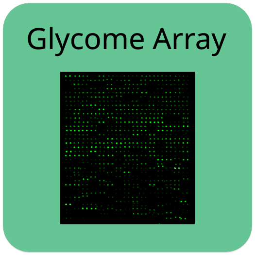
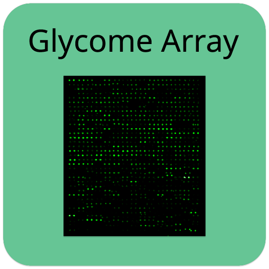
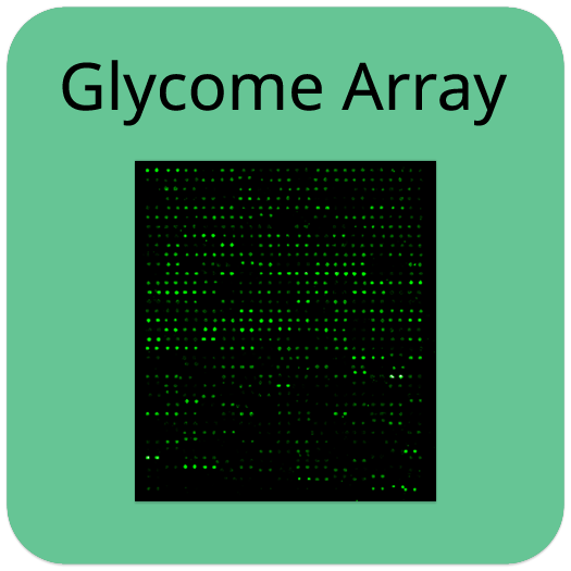
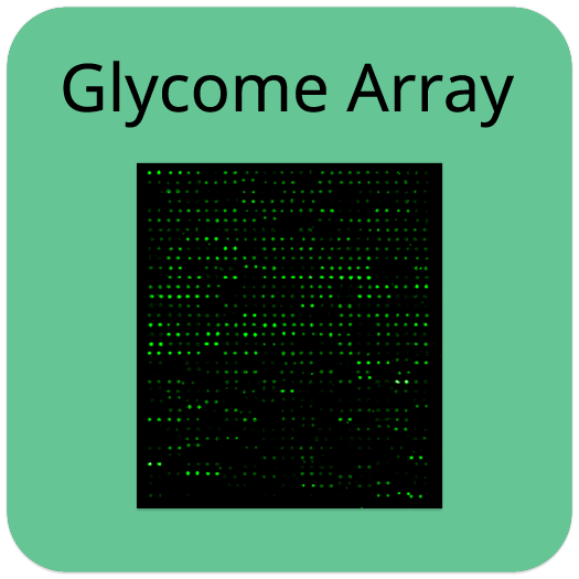
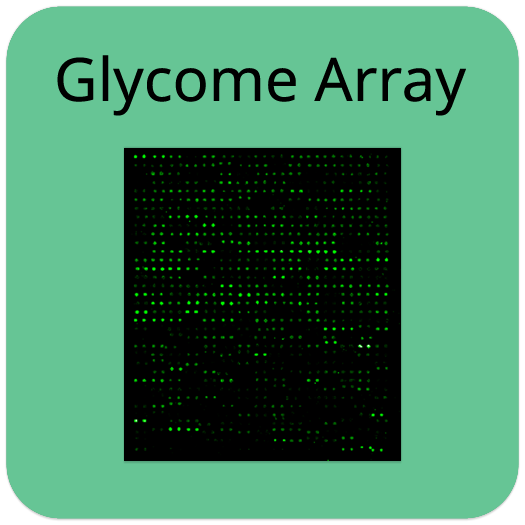
The following figure shows the Human Glycosylation Antibody Array 507 probed with conditioned media sample. The images were captured using a Axon GenePix laser scanner. The strong signals in row 20 and the upper left and lower right corners of each array are Positive Controls, which can be used to identify the orientation and help normalize the results between arrays.
Suggested Applications
Identify and profile the glycans in their samples
Determine whether their biomarker of interest has glycan moieties
Determine relative expression levels
Kit Components
Human Glycosylation Array Glass Slides
Sample Diluent
20X Wash Buffer I
20X Wash Buffer II
Cy3 equivalent dye-conjugated Streptavidin
Biotin-labeled Lectin Mixture
Adhesive device sealer
Manual
Other Materials Required
Pipettors, pipet tips and other common lab consumables
Orbital shaker or oscillating rocker
Aluminum foil
Distilled or de-ionized water
1 ml microcentrifuge tubes
Gene microarray scanner or similar laser fluorescence scanner
Protocol Outline
Dry the glass slide
Incubate samples
Incubate with biotin lectin mixture
Incubate with Cy3 Equivalent Dye-Streptavidin
Disassemble the glass slide
Scan with a gene microarray laser scanner
Perform densitometry and analysis
Storage/Stability
Upon receipt, all components of the Human Glycosylation Array 1000 kit should be stored at -20 ° C. Once thawed, the glass slide and Cy3 equivalent dye-conjugated Streptavidin should be kept at -20 ° C and all other components may be stored at 4 ° C. The entire kit should be used within 6 months of purchase.