 from Microorganism")
| Appearance | White amorphous powder, lyophilized | |
|---|---|---|
| Activity | GradeⅡ 20 U/mg-solid or more | |
| Contaminants | α-amylase | ≤1.0×10⁻⁵ % |
| Stabilizers | BSA | |
| Stability | Stable at −20 ℃ for at least one year(Fig.1) | ||
|---|---|---|---|
| Molecular weight | approx. 65,000 (Gel-filtration and SDS-PAGE) | ||
| Isoelectric point | 5.2 | ||
| Michaelis constant | 6.3×10⁻⁴M (p-Nitrophenyl-α-D-glucopyranoside) | ||
| Inhibitors | Ag+, Hg2+, PCMB, MIA | ||
| Optimum pH | 6.0−7.0(Fig.4) | ||
| Optimum temperature | 60 ℃(Fig.5) | ||
| pH Stability | pH 5.0−9.0(Fig.6) | ||
| Effect of various chemicals | (Table 1) | ||
| Thermal stability | below 60℃ (pH 7.0, 15min)(Fig.7) | ||
| Substrate* | Relative hydrolysis rate** | Substrate* | Relative hydrolysis rate** |
|---|---|---|---|
| PNPG | 100.0 | Maltose | 271.0 |
| PNPG2 | 205.0 | Maltotriose | 203.0 |
| PNPG3 | 284.0 | Maltotetraose | 168.0 |
| PNPG5 | 164.0 | Maltopentaose | 100.0 |
* : Substrate concn. 2.2mM
** : Glucose-forming activity, pH 6.8 at 37℃
This enzyme is useful for structural investigation of carbohydrates and for enzymatic determination of α-amylase when in combination hexokinase (HXK-311) and G-6-P dehydrogenase (G6D-311, G6D321) in clinical analysis.
![]()
The formation of p-nitrophenol is measured at 400 nm by spectrophotometry.
One unit causes the formation of one micromole of PNP per minute under the conditions detailed below.
| A. 0.1M Phosphate buffer, pH 7.0 (at 25℃) | |
|---|---|
| B. PNPG solution | 20 mM (603mg P-Nitrophenyl-α-D-glucopyranoside /100 mL of H2O)(Stable for two weeks if stored at 0−5℃) |
| C. Na2CO3 solution | 0.2 M (21.2g Na2CO3 /1,000 mL of H2O) |
| D. Enzyme diluent | 0.2 M K-phosphate buffer, pH 7.0 containing 1mM of EDTA and 0.05 % of Tween 20 |
1.Prepare the following reaction mixture in a test tube and equilibrate at 37℃ for approximately 5 minutes.
| 1.0 mL | 0.1 M phosphate buffer | (A) |
| 0.5 mL | Substrate solution | (B) |
| Concentration in assay mixture | |
|---|---|
| Phosphate buffer | 0.1 M |
| PNPG | 5.0 mM |
| EDTA | 0.25 mM |
| Tween 20 | 0.125 mg/mL |
2.Add 0.5 mL of the enzyme solution* and mix.
3.After exactly 15 minutes at 37℃, add 2.0 mL of Na2CO3 solution (C) to stop the reaction and measure the optical density at 400nm against water (OD test).
At the same time, prepare the blank by mixing the reaction mixture with 2.0 mL of Na2CO3 solution (C) after incubation for 15 minutes at 37℃, followed by the addition of the enzyme solution (OD blank).
*Dissolve the enzyme preparation in ice-cold enzyme diluent (D) and dilute to 0.006−0.022U/mL with the same buffer, immediately before the assay.
Activity can be calculated by using the following formula :
Volume activity (U/mL) =
ΔOD (OD test−OD blank)×Vt×df
18.1×t×1.0×Vs
= ΔOD×0.0295×df
Weight activity (U/mg) = (U/mL)×1/C
| Vt | : Total volume (4.0 mL) |
| Vs | : Sample volume (0.5 mL) |
| 18.1 | : Millimolar extinction coefficient of p-nitrophenol under the assay condition (cm2/micromole) |
| 1.0 | : Light path length (cm) |
| t | : Reaction time (15 minutes) |
| df | : Dilution factor |
| C | : Enzyme concentration in dissolution (c mg/mL) |
1)Y.Suzuki, M.Shinji and N.Eto; Biochim.Biophys.Acta., 787, 281 (1984).
2)Y.Takii, K.Daimon and Y.Suzuki; Appl.Microbiol.Biotechnol., 38, 243 (1992).
3)Y.Takii, K.Takahashi, K.Yamamoto, Y.Sogabe and Y.Suzuki; Appl.Microbiol.Biotechnol., 44, 629 (1996).
[The enzyme dissolved in 10mM phosphate buffer, pH 7.0 contg. 0.2 % of BSA (5 U/mL) was incubated with each chemical at 25 ℃ for 1 hr.]
| Chemical | Concn.(mM) | Residual activity(%) |
|---|---|---|
| None | – | 100 |
| Metal salt | 2.0 | |
| MgSO4 | 97 | |
| CaCl2 | 71 | |
| Ba(OAc)2 | 106 | |
| FeCl2 | 50 | |
| CoCl2 | 63 | |
| MnCl2 | 69 | |
| ZnCl2 | 104 | |
| CdCl2 | 47 | |
| NiCl2 | 110 | |
| CuSO4 | 39 | |
| Pb(OAc)2 | 75 | |
| AgNO2 | 0.3 | |
| HgCl2 | 1.2 | |
| 2-Mercaptoethanol | 2.0 | 111 |
| PCMB | 1.0 | 1.3 |
| Chemical | Concn.(mM) | Residual activity(%) |
|---|---|---|
| MIA | 2.0 | 0.8 |
| NEM | 2.0 | 120 |
| IAA | 2.0 | 106 |
| Hydroxylamine | 2.0 | 115 |
| EDTA | 5.0 | 112 |
| o-Phenanthroline | 2.0 | 114 |
| α,α′-Dipyridyl | 1.0 | 122 |
| Borate | 50 | 119 |
| NaF | 2.0 | 118 |
| NaN3 | 2.0 | 123 |
| Triton X-100 | 0.10 % | 123 |
| Brij 35 | 0.10 % | 121 |
| Tween 20 | 0.10 % | 124 |
| Span 20 | 0.10 % | 43 |
| Na-cholate | 0.10 % | 102 |
| SDS | 0.05 % | 10 |
| DAC | 0.05 % | 124 |
Ac, CH3CO; PCMB, p-Chloromercuribenzoate; MIA, Monoiodoacetate; NEM, N-Ethylmaleimide; IAA, Iodoacetamide; EDTA, Ethylenediaminetetraacetate; SDS, Sodium dodecyl sulfate; DAC, Dimethylbenzylalkylammonium chloride.
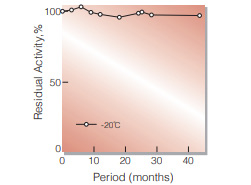
Fig.1. Stability (Powder form)
(kept under dry conditions)
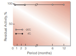
Fig.2. Stability (Powder form)
(kept under dry conditions)
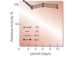
Fig.3. Stability (Liquid form)
in 50mM PIPES buffer solution, pH7.0 (contg. 0.5mM CaCl2, 0.1 % detergent) enzyme concn,:5U/mL
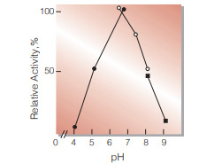
Fig.4. pH-Activity
37℃, 15 min-reaction in 100mM buffer solution: ●, pH4.0-6.0 acetate ; O, pH6.0-8.0, phosphate; ■, pH8.0-9.0, borate
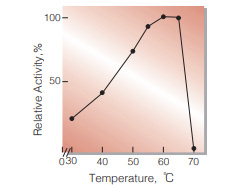
Fig.5. Thermal activity
15 min-reaction in 100mM phosphate buffer, pH7.0
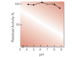
Fig.6. pH-Stability
25℃, 20hr-treatment with 50mM buffer solution contg; 0.2 % of BSA: ●, pH4.0-6.0 acetate: ○, pH6.0-8.0, phosphate; ■, pH8.0-9.0, borate. enzyme concn. : 5U/mL
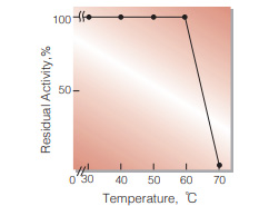
Fig.7. Thermal stability
15min-treatment with 0.2M K-phosphate buffer, pH7.0 contg. 1mM EDTA and 0.05 % Tween20. enzyme concn.: 5U/mL
| Size | 1 MG, 10 MG, 5 MG |
|---|
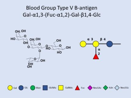
Gal-a1,3-(Fuc-a1,2)-Gal-b1,4-Glc, CAS:59957-92-5, G06873PC
10 in stock
10 in stock
GalNAc-a1,3-(Fuc-a1,2)-Gal-b1,4-Glc, CAS:59957-92-5, G19524AS
10 in stock
10 in stock

Reviews
There are no reviews yet.