 from Microorganism")
| Appearance | Purple amorphous powder, lyophilized | |
|---|---|---|
| Activity | Grade Ⅲ 500 U/mg-solid or more | |
| Contaminants | Glucose dehydrogenase (NAD-dependent) | ≤ 1.0×10⁻³ % |
| Hexokinase | ≤ 1.0×10⁻³ % | |
| Stabilizers | Ca2+, BSA | |
| Stability | Stable at − 20℃ for at least one year(Fig.1) |
|---|---|
| Molecular weight | approx. 100,000 (by gel filtration) |
| Michaelis constant | 4.8 mM (D-Glucose) |
| Inhibitors | Cu2+, Pb2+, Ag+ |
| Optimum pH | 7.0(Fig.2) |
| Optimum temperature | 37 ℃(Fig.3) |
| pH Stability | pH 3.5−8.5 (25 ℃, 16 hr)(Fig.4) |
| Thermal stability | below 50 ℃ (pH 7.5, 30 min)(Fig.5) |
| Substrate specificity | (Table 1) |
| Effect of various chemicals | (Table 2) |
This enzyme is useful for enzymatic determination of D-glucose.
![]()
The formation of diformazan formed by the reduction of nitrotetrazorium blue (NTB) with phenazine methosulfate (PMS), which is red, is measured at 570 nm by spectrophotometry.
One unit causes the formation of one half micromole of diformazan per minute under the conditions detailed below.
| A. D-Glucose solution | 1 M: 1.8 g of D-glucose (MW = 180.16) / 10 mL of H2O (keep this solution at room temperature for at least 3 hours before use). |
|---|---|
| B. PIPES-NaOH buffer, pH 6.5 | 50 mM: Weigh out 1.51 g of PIPES (MW = 302.36), suspend in 60 mL of H2O, dissolve with 5 N NaOH, and add 2.2 mL of 10 % Triton X-100. After adjusting pH to 6.5 ± 0.05 at 25℃ with 5 N NaOH, make up to 100mL with H2O. |
| C. PMS solution | 3.0 mM: 9.19 mg of phenazine methosulfate (MW = 306.34) / 10 mL of H2O |
| D. NTB solution | 6.6 mM: 53.96 mg of NTB (MW = 817.65) / 10 mL of H2O |
| E. Enzyme diluent | 50 mM PIPES-NaOH buffer, pH 6.5, containing 1 mM CaCl2, 0.1 % Triton X-100, 0.1 % BSA |
1.Prepare the following reaction mixture in a brownish bottle shortly before use, and store on ice.
| 0.9 mL | D-glucose solution | (A) |
| 25.5 mL | PIPES-NaOH buffer, pH 6.5 | (B) |
| 2.0 mL | PMS solution | (C) |
| 1.0 mL | NTB solution | (D) |
| Concentration in assay mixture | |
|---|---|
| PIPES-buffer | 42 mM |
| D-glucose | 30 mM |
| PMS | 0.20 mM |
| NTB | 0.22 mM |
2.Pipette 3.0 mL of working solution into a plastic test tube and equilibrate at 37 ℃ for approximately 5 minutes.
3.Add 0.1 mL of enzyme solution* and mix by gentle inversion.
4.Record the increase of optical density at 570 nm against water for 4 to 5 minutes with a spectrophotometer thermostated at 37 ℃, and calculate the ΔOD per minute from the initial linear portion of the curve (ΔOD test).
At the same time, measure the blank rate (ΔOD blank) by the same method as in the test except that the enzyme diluent (E) is added instead of the enzyme solution.
*Dissolve the enzyme preparation on ice cold enzyme diluent (E) and dilute to 0.1−0.8 U/mL with the same buffer, immediately before the assay. (A plastic tube is recommended because of viscous properties of the liquid.)
Activity can be calculated by using the following formula :
Volume activity (U/mL) =
ΔOD/min (ΔOD test−ΔOD blank)×Vt×df
20.1×1.0×Vs
= ΔOD×1.54×df
Weight activity (U/mg) = (U/mL)×1/C
| Vt | : Total volume (3.1 mL) |
| Vs | : Sample volume (0.1 mL) |
| 20.1 | : Half a millimolar extinction coefficient of diformazan (cm2/0.5 micromole) |
| 1.0 | : Light path length (cm) |
| df | : Dilution factor |
| C | : Enzyme concentration in dissolution (c mg/mL) |
1)K.Matsushita et al.; FEMS Microbiology Letters, 55, 53 (1988).
| Substrate (50mM) | Relative activity(%) |
|---|---|
| D-Glucose | 100.0 |
| L-Glucose | 0.3 |
| D-Xylose | 15.0 |
| 2-Deoxy-glucose | 4.9 |
| L-Sorbose | 0.5 |
| D-Mannose | 10.8 |
| D-Fructose | 0.3 |
| Substrate (50mM) | Relative activity(%) |
|---|---|
| Galactose | 16.0 |
| D-Lactose | 68.9 |
| D-Sorbitole | 0.2 |
| D-Mannitol | 0.0 |
| Sucrose | 0.2 |
| Inositol | 0.0 |
| Maltose | 107.0 |
[The enzyme dissolved in 50mM PIPES-NaOH buffer, pH 6.5 contg. 1mM CaCl2, 0.1% Triton X-100 (5U/mL) was incubated with each chemical at 25℃ for 1hr.]
| Chemical | Concn.(mM) | Residual activity(%) |
|---|---|---|
| None | – | 100 |
| Metal salt | 2.0 | |
| MgSO4 | 108 | |
| CaCl2 | 108 | |
| Ba(OAc)2 | 105 | |
| FeCl3 | 79 | |
| CoCl2 | 42 | |
| MnCl2 | 105 | |
| ZnCl2 | 45 | |
| Cd(OAc)2 | 107 | |
| NiCl2 | 101 | |
| CuSO4 | 0 | |
| Pb(OAc)2 | 0 | |
| AgNO3 | 0 | |
| HgCl2 | 77 | |
| 2-Mercaptoethanol | 2.0 | 99 |
| PCMB | 1.0 | 97 |
| Chemical | Concn.(mM) | Residual activity(%) |
|---|---|---|
| MIA | 2.0 | 87 |
| NEM | 2.0 | 100 |
| IAA | 2.0 | 98 |
| Hydroxylamine | 2.0 | 19 |
| EDTA | 5.0 | 79 |
| O-Phenanthroline | 2.0 | 7 |
| α,α′-Dipyridyl | 1.0 | 103 |
| Borate | 5.0 | 110 |
| NAF | 2.0 | 111 |
| NaN3 | 2.0 | 115 |
| Triton X-100 | 0.10 % | 101 |
| Brij 35 | 0.10 % | 22 |
| Tween 20 | 0.10 % | 104 |
| Span 20 | 0.10 % | 60 |
| Na-Cholate | 0.10 % | 67 |
| SDS | 0.05 % | 33 |
| DAC | 0.05 % | 113 |
Ac, CH3CO; PCMB, p-Chloromercuribenzoate; MIA, Monoiodoacetate; NEM, N-Ethylmaleimide; IAA, Iodoacetamide; EDTA, Ethylenediaminetetraacetate; SDS, Sodium dodecyl sulfate; DAC, Dimethylbenzylalkylammonium chloride.
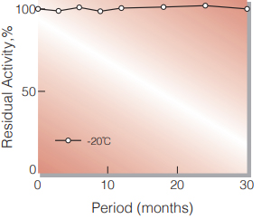
Fig.1. Stability (Powder form)
(kept under dry conditions)
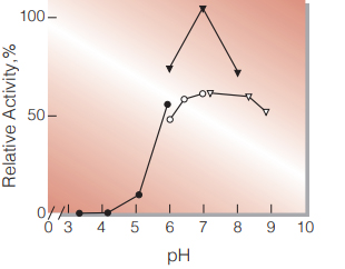
Fig.2. pH-Activity
25℃, in 50mM buffer solution; ●̶●,acetate; ▼̶▼, phosphate; ○̶○, PIPES; ▽̶▽, Tris-HCl.
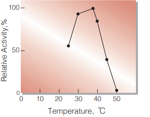
Fig.3. Temperature Activity
(in 42mM PIPES-NaOH buffer, pH 6.5)
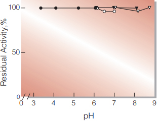
Fig.4. pH-Stability
25℃, 16 hr-treatment with 50mM buffer solution contg. 1mM CaCl2; ●̶●,acetate; ▼̶▼, phosphate; ○̶○, PIPES; ▽̶▽, Tris-HCl.
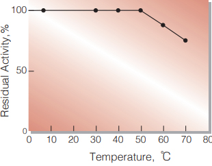
Fig.5. Thermal stability
30min.-treatment with 50mM PIPES-NaOH buffer, pH 6.5 contg. 1mM CaCl2 enzyme concentration: 5.0 U/mL
| Size | 1 MG, 10 MG, 5 MG |
|---|
GalNAc-a1,3-(Fuc-a1,2)-Gal-b1,4-Glc, CAS:59957-92-5, G19524AS
10 in stock
10 in stock
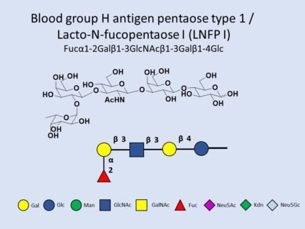
IUPAC:Fuc(a1-2)Gal(b1-3)GlcNAc(b1-3)Gal(b1-4)Glc, G00050MO
10 in stock
Catalog No: CG-OS03017
CAS No: 14116-68-8
Synonyms: Galβ1-3GlcNAcβ1-3Galβ1-4Glc; LNT
Molecular Formula: C₂₆H₄₅NO₂₁
Molecular Weight: ~707.63 g/mol
Available Pack Sizes: Research quantities (mg to g scale)
10 in stock


Reviews
There are no reviews yet.