
GLUCOSE-6-PHOSPHATE DEHYDROGENASE from Leuconostoc mesenteroides
$100.10 – $200.10
| Appearance : | White amorphous powder, lyophilized | |
|---|---|---|
| Activity : | GradeⅢ 400 U/mg-solid or more (NAD+) | |
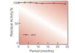
| Stability : | Stable at −20 ℃ for at least one year | |
|---|---|---|
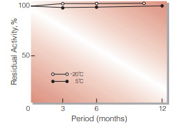
| Stable at 5 ℃ for at least 6 months (liquid form) | ||
| Molecular weight : | 104,000(two subunits of approx. 55,000) 1,2) | |
PREPARATION and SPECIFICATION
| Appearance | White amorphous powder, lyophilized | |
|---|---|---|
| Activity | GradeⅢ 400 U/mg-solid or more (NAD+) | |
| Contaminants | Creatine phosphokinase | ≤ 1×10⁻³ % |
| Phosphoglucomutase | ≤ 1×10⁻³ % | |
| 6-Phosphogluconate dehydrogenase | ≤ 5×10⁻³ % | |
| Phosphoglucose isomerase | ≤ 1×10⁻² % | |
| Glutathione reductase | ≤ 1×10⁻³ % | |
| Hexokinase | ≤ 1×10⁻² % | |
| Myokinase | ≤ 1×10⁻² % | |
| NADH oxidase | ≤ 1×10⁻² % | |
| NADPH oxidase | ≤ 1×10⁻² % | |
PROPERTIES
| Stability | Stable at −20 ℃ for at least one year(Fig.1) | |
|---|---|---|
| Stable at 5 ℃ for at least 6 months (liquid form)(Fig.3) | ||
| Molecular weight | 104,000(two subunits of approx. 55,000) 1,2) | |
| Isoelectric point | 4.6 2) | |
| Michaelis constants 2) | NAD+ linked | 1.06×10⁻⁴ M (NAD+), 5.27×10⁻⁵ M (G-6-P) |
| NADP+ linked | 5.69×10⁻⁶ M (NADP+), 8.1×10⁻⁵ M (G-6-P) | |
| Structure | Neither cysteine nor cystine residues is present in the enzyme molecule 1) and essential lysine is indicated to be at active site. 3) | |
| Inhibitors | Acyl-CoA,4) ATP,4) mental ions etc. (Table 1) | |
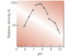
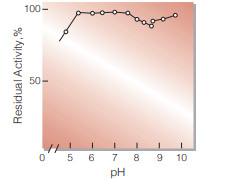
| Optimum pH | 7.8(Fig.4) | |
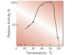
| Optimum temperature | 50 ℃(Fig.5) | |
| pH Stability | pH 5.5−7.5 (30 ℃, 17 hr)(Fig.6) | |
| Thermal stability | below 37 ℃ (pH 8.0, 30 min)(Fig.7) | |
| Substrate specificity | Either NAD+ or NADP+ serves as coenzyme, the reaction velocity with NAD+ being approximately 1.8 times greater than with NADP.+5) DGlucose-6-phosphate is a preferential substrate for the enzyme, although D-glucose reacts slowly.6) Fructose-6-phosphate, fructose1, 6-diphoshate and ribose-5-phosphate are not considered to be substrates.7) | |
APPLICATIONS
This enzyme is useful for enzymatic determation of NAD+(NADP+) and G-6-P, and the activities of phosphoglucose isomerase, phosphoglucomutase and hexokinase. This enzyme is also used for enzymatic determination of glucose in combination with hexokinase (HXK-311).
ASSAY
Principle
![]()
The formation of NADH is measured at 340 nm by spectrophotometry.
Unit definition
One unit causes the formation of one micromole of NADH per minute under the conditions detailed below.
Method
Reagents
| A. Tris-HCl buffer, pH 7.8 | 55 mM (containing 3.3 mM magnesium chloride) | |
|---|---|---|
| B. NAD+ solution | 60 mM (Should be prepared fresh) | |
| C. G-6-P solution | 0.1 M glucose-6-phosphate (should be prepared fresh) | |
| D. Enzyme diluent | 5 mM Tris-HCl buffer, pH 7.5, containing 0.1 % of bovine serum albumin. | |
Procedure
1.Prepare the following reaction mixture in a cuvette (d = 1.0 cm) and equilibrate at 30 ℃ for about 5 minutes.
| 2.7 mL | Tris-HCl buffer, pH 7.8 | (A) |
| 0.1 mL | NAD+ solution | (B) |
| 0.1 mL | G・6・P solution | (C) |
| Concentration in assay mixture | |
|---|---|
| Tris-HCl buffer | 50 mM |
| G-6-P | 3.3 mM |
| NAD+ | 2.0 mM |
| MgCl2 | 3.0 mM |
| BSA | 33 μg/mL |
2.Add 0.1 mL of the enzyme solution* and mix by gentle inversion.
3.Record the increase in optical density at 340 nm against water for 4 to 5 minutes with a spectrophotometer thermostated at 30 ℃ and calculate the ΔOD per minute from the initial linear portion of the curve (ΔOD test).
At the same time, measure the blank rate (ΔOD blank) using the same method as the test except that the enzyme diluent is added instead of the enzyme solution.
*Dissolve the enzyme preparation in ice-cold enzyme diluent (D) and dilute to 0.05−0.20 U/mL with the same buffer, immediately before the assay.
Calculation
Activity can be calculated by using the following formula :
Volume activity (U/mL) =
ΔOD/min (ΔOD test−ΔOD blank)×Vt×df
6.22×1.0×Vs
= ΔOD/min×4.82×df
Weight activity (U/mg) = (U/mL)×1/C
| Vt | : Total volume (3.0 mL) |
| Vs | : Sample volume (0.1 mL) |
| 6.22 | : Millimolar extinction coefficient of NADH (cm2/micromole) |
| 1.0 | : Light path length (cm) |
| df | : Dilution facter |
| C | : Enzyme concentration in dissolution (c mg/mL) |
REFERENCES
1)A.Ishaque,M.Mihausen and H.R.Levy; Biochem. Biophys. Res. Comm., 59, 894 (1974).
2)C. Olive, M.E. Geroch and H.R.Levy; J.Biol.Chem., 246, 2043 (1971).
3)M.Milhausen and H.R. Levy; Eur.J.Biochem., 50, 453 (1975).
4)E.L.Coe and L.-H.Hsu; Biochem. Biophys. Res. Comm., 53, 66 (1973).
5)C.Olive and H.R. Levy; Biochem., 6, 730 (1967).
6)R.P.Metzger, S.A. Metzger and R.L. Parsons; Arch Biochem. Biophys., 149, 102 (1972).
7)Methods in Enzymology, Vol, 1, p328 (S.P.Colowick and N.O.Kapalan,eds.), Academic Press, New York (1955).
Table 1. Effect of Various Chemicals on Glucose-6-phosphate dehydrogenase
[The enzyme dissolved in 50 mM Tris-HCl buffer,pH 7.5 (5.25 U/mL) was incubated with each chemcal for 1 hr at 30 ℃.]
Chemical Concn.(mM) Residual
activity(%)None – 100 Metal salt 2.0 AgNO3 86 Ba(OAc)2 51 CaCl2 90 Cd(OAc)2 74 CoCl2 80 CuSO4 66 FeCl3 0 FeSO4 1 HgCl2 84 MgCl2 90 MnCl2 89 NiCl2 89 Pb(OAc)2 3 Zn(OAc)2 67 ZnSO4 53 KF 2.0 93 NaF 20.0 98 NaN3 20.0 93 Chemical Concn.(mM) Residual
activity(%)NEM 2.0 91 PCMB 2.0 96 MIA 2.0 14 Iodoacetamide 2.0 0 EDTA 5.0 94 (NH4)2SO4 20.0 98 Borate 20.0 95 o-Phenanthroline 2.0 93 α,α′-Dipyridyl 2.0 95 Urea 2.0 93 Guanidine 2.0 93 Hydroxylamine 2.0 91 Na-cholate 1.0 % 102 Triton X-100 1.0 % 100 Brij 35 1.0 % 4 SDS 0.1 % 0 Tween 20 0.1 % 101 Span 20 0.1 % 99 DAC 0.1 % 0
Ac, CH3CO; NEM, N-Ethylmaleimide; PCMB, p-Chloromercuribenzoate; MIA, Monoiodoacetate; EDTA, Ethylenediaminetetraacetate; SDS, Sodium dodecyl sulfate; DAC, Dimethylbenzylalkylammoniumchloride.

Fig.1. Stability (Powder form)
(kept under dry conditions)
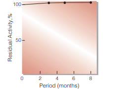
Fig.2. Stability (Powder form)
(kept under dry conditions)
Fig.3. Stability (Liquid form at 5 ℃)
enzyme concentration:5,000 U/mL composition:3.2 M ammonium sulfate,pH 6.0
Fig.4. pH-Activity
30 ℃ in the following buffer solution: pH 5.7-6.8, 15 mM Veronal-CH3COONaHCI;pH 6.8-8.5,50 mM Tris-HCI; pH 8.5-9.5, 50 mM glycine-NaOH
Fig.5. Temperature activity
(in 50 mM Tris-HCI buffer, pH 7.8)
Fig.6. pH-Stability
30 ℃, 17 hr-treatment with the following buffer solution: pH 5.0-7.8, 30 mM VeronalCH3COONa-HCI;pH 7.5-8.5, 0.1 M Tris-HCI; pH 8.5-9.5,0.1 M glycine-NaOH
Fig.7. Thermal stability
30 min-treatment with 5.0 mM glycineNaOH buffer, pH 8.0, containing 0.1 % of bovine serum albumin

| Size | 1 MG, 10 MG, 5 MG |
|---|
Related Products

| Name | β1,4-galactosyltransferase; LgtB |
| Catalog Number | EN01005 |
| E.C. | 2.4.1.90 |
| Product Description | E. coli recombinant β1,4-galactosyltransferase from Neisseria meningitides |
| Unit Definition | One unit is defined as the amount of enzyme that catalyzes the formation of 1 µmol of Galβ1,4GlcNAc from UDP-Gal and GlcNAc per min at 37 °C. |
10 in stock

| Name | GlcNAc-1-P uridyltransferase (GlmU) |
| Catalog No. | EN01012 |
| E.C. | 2.3.1.157 |
10 in stock

| Name | L-fucokinase-GDP-fucose pyrophos-phorylase;FKP |
| Product Code | EN01016 |
| E.C. | 2.7.1.52/2.7.7.30 |
| Product Description | E. coli recombinant L-fucokinase/GDP-fucose pyrophosphorylase from Bacteroides fragilis |
| Unit Definition | One unit is defined as the amount of enzyme that catalyzes the formation of 1 μmol of Fuc-1-P from L-Fuc and ATP per minute at 37 °C. |
10 in stock

| Name | Sialic acid aldolase (NPL) |
| Catalog No. | EN01027 |
| E.C. | 4.1.3.3 |
10 in stock
Reviews
There are no reviews yet.