


From: $5,583.00
| Size | 4 Sample Kit, 8 Sample Kit |
|---|---|
| Species | Human |
| Quantitative/Semi-Quantitative | Semi-Quantitative |
| Number of Targets Detected | 8000 |
| Compatible Sample Types | Cell Culture Supernatants, Plasma, Serum, Tissue Lysates, Cell Lysates |
| Solid Support | Glass Slide |
| Method Of Detection | Fluorescence Laser Scanner |
| Design Principle | Sandwich-based |
| Research Area | Post-Translational Modifications, Glycosylation |
| Estimated Lead Time | 1-2 business days |
| Shipping Type | Blue ice |
| Storage | -20°C |
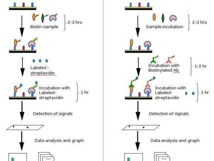
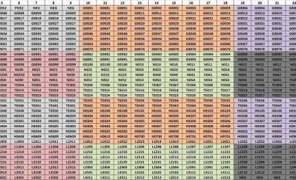
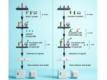
Capture antibodies are printed onto glass slides and the glycans on these capture antibodies are removed. The glass slide arrays come pre-blocked and are ready to be incubated with samples. After incubation with samples and washing to remove unbound proteins, five unique biotin-labeled lectins are incubated with the array to allow binding to the glycans from captured target proteins. Streptavidin-conjugated fluorescent dye (Cy3 equivalent) is then added to the array. Finally, the glass slide is dried and laser fluorescence scanning is used to visualize the signals. These signals are then compared to the array map to identify glycosylated proteins present in the samples.
Biotin-Labeled Lectin Mixture | ||
Lectin Name | Sugar specificity | |
1 | Concanavalin A | αMan, αGlc |
2 | Dolichos Biflorus Agglutinin | αGalNAc |
3 | Peanut Agglutinin | Galβ3GalNAc |
4 | Ulex Europaeus Lectin 1 | αFuc |
5 | Wheat Germ Agglutinin | GlcNAc |

| sample size | 4 Sample Kit, 8 Sample Kit |
|---|
| Size | 4 Sample Kit, 8 Sample Kit |
|---|---|
| Species | Human |
| Quantitative/Semi-Quantitative | Semi-Quantitative |
| Number of Targets Detected | 1000 |
| Compatible Sample Types | Cell Culture Supernatants, Plasma, Serum, Tissue Lysates, Cell Lysates |
| Solid Support | Glass Slide |
| Method Of Detection | Fluorescence Laser Scanner |
| Design Principle | Sandwich-based |
| Research Area | Post-Translational Modifications, Glycosylation |
| Estimated Lead Time | 1-2 business days |
| Shipping Type | Blue ice |
| Storage | -20°C |
| Size | 4 Sample Kit, 8 Sample Kit |
|---|---|
| Species | Human |
| Quantitative/Semi-Quantitative | Semi-Quantitative |
| Number of Targets Detected | 500 |
| Compatible Sample Types | Cell Culture Supernatants, Plasma, Serum, Tissue Lysates, Cell Lysates |
| Solid Support | Glass Slide |
| Method Of Detection | Fluorescence Laser Scanner |
| Design Principle | Sandwich-based |
| Research Area | Post-Translational Modifications, Glycosylation |
| Estimated Lead Time | 1-2 business days |
| Shipping Type | Blue ice |
| Storage | -20°C |
| Size | 12 Sample Kit, 24 Sample Kit, 48 Sample Kit |
|---|---|
| Quantitative/Semi-Quantitative | Semi-Quantitative |
| Number of Targets Detected | 95 |
| Compatible Sample Types | Cell Culture Supernatants, Plasma, Serum, Tissue Lysates, Cell Lysates |
| Solid Support | Glass Slide |
| Method Of Detection | Fluorescence Laser Scanner |
| Design Principle | Sandwich-based, Label-based |
| Research Area | Post-Translational Modifications, Glycosylation |
| Estimated Lead Time | 1-2 business days |
| Shipping Type | Blue ice |
| Storage | -20°C |
| Size | 4 Sample Kit, 8 Sample Kit, 16 Sample Kit |
|---|---|
| Quantitative/Semi-Quantitative | Semi-Quantitative |
| Number of Targets Detected | 300 |
| Compatible Sample Types | Cell Culture Supernatants, Plasma, Serum, Tissue Lysates, Cell Lysates, Purified Proteins |
| Solid Support | Glass Slide |
| Method Of Detection | Fluorescence Laser Scanner |
| Design Principle | Sandwich-based, Label-based |
| Research Area | Post-Translational Modifications, Glycosylation |
| Estimated Lead Time | 1-2 business days |
| Shipping Type | Blue ice |
| Storage | -20°C |


Reviews
There are no reviews yet.