
| Appearance | White amorphous powder, lyophilized. | |
|---|---|---|
| Activity | GradeⅡ 500 U/mg-solid or more | |
| Contaminants | α-galactosidase | < 1×10⁻⁴ % |
| α-glucosidase | < 1×10⁻⁴ % | |
| β-glucosidase | < 2×10⁻³ % | |
| α-mannosidase | < 1×10⁻⁴ % | |
| β-mannosidase | < 1×10⁻⁴ % | |
| proteinasee | <10mAbs/mg-P | |
| Stabilizer | Mg2+ | |
| Stability | Stable at −20 ℃ for at least one year(Fig.1) |
|---|---|
| Molecular weight | 540,000 1,2) |
| Isoelectric point 3) | 4.6 |
| Michaelis constants | 3.0×10⁻⁴ M (o-Nitrophenyl-β-D-galactoside), 6.7×10⁻⁵ M (pNitrophenyl-β-D-galactoside), 2.3×10⁻⁴ M (Phenyl-β-D-galactoside), 2.5×10⁻³ M (Lactose) |
| Structure 4〜8) | The enzyme is composed of four identical subunits having a molecular weight of ca.135,000. The amino acid analysis indicates approximately 1,170 residues per subunit. E 280 nm1 cm (1%)=20.9 1 cm |
| Inhibitors | p-Chloromercuribenzoate, lodoacetamide, heavy metal ions (Zn2+, Fe3+, Cd2+, Cu2+, Pb2+, Ag+, Hg2+), lonic detergents (SDS, DAC, etc.) |
| Optimum pH | 7.0−7.5(Fig.2) |
| Optimum temperature | 50−55 ℃(Fig.3) |
| pH Stability | pH 6.5−8.5 (25 ℃, 20 hr)(Fig.4) |
| Thermal stability | below 50 ℃ (pH 7.3, 15 min)(Fig.5) |
| Substrate specificty | This enzyme specifically hydrolyzes β-D-galactosyl linkage (Table 1). |
| Effect of various chemicals | (Table 2) |
This enzyme is useful for structural analysis of carbohydrates, measurement of lactose (foodstuff analysis) and as an enzyme label for enzyme immunoassays.
![]()
The formation of o-nitrophenol is measured at 410 nm by spectrophotometry.
One unit causes the formation of one micromole of ONP per minute under the conditions detailed below.
| A. Phosphate buffer, pH 7.3 | 0.1 M: Prepare by mixing 0.1 M Na2HPO4 and 0.1 M KH2PO4 to pH 7.3 at 37 ℃.) |
|---|---|
| B. Mercaptoethanol solution | 3.36 M: Dilute 4.0 mL of 2-mercaptoethanol (14.2 M) to 17 mL with H2O (should be freshly prepared). |
| C. MgCl2 solution | 30 mM: Dissolve 610mg of MgCl2・6H2O in approx. 80 mL of H2O and, after adjusting the pH to 7.3 with 1.0 NaOH, fill up to 100 mL with H2O. |
| D. ONPG solution | 34 mM: 205 mg of ONPG / 20 mL of reagent A (stable for 1 week if stored at 0−5 ℃). |
| E. Enzyme diluent | 50mM phosphate buffer, pH 7.3, containing 1.0mM MgCl2 and 0.1 % BSA |
1.Prepare the following reaction mixture in a cuvette (d = 1.0cm) and equilibrate at 37℃ for approximately 5 minutes.
| 2.5 mL | 0.1M Phosphate buffer, pH 7.3 | (A) |
| 0.1 mL | Mercaptoethanol solution | (B) |
| 0.1 mL | MgCl2 solution | (C) |
| 0.2 mL | ONPG solution | (D) |
| Concentration in assay mixture | |
|---|---|
| Phosphate buffer | 92 mM |
| ONPG | 2.3 mM |
| Mercaptoethanol | 0.11 M |
| MgCl2 | 1.0 mM |
2.Add 0.1 mL of the enzyme solution* and mix by gentle inversion
3.Record the increase in optical density at 410 nm against water for 2 to 3 minutes with a spectrophotometer thermostated at 37 ℃, and calculate ΔOD per minute from the initial linear portion of the curve (ΔOD test).
At the same time, measure the blank rate (ΔOD blank) using the same method as the test except that the enzyme diluent (E) is added instead of the enzyme solution.
*Dilute the enzyme preparation to 0.17−0.85 U/mL with ice-cold enzyme diluent (E).
Activity can be calculated by using the following formula :
Volume activity (U/mL) =
ΔOD/min (ΔOD test−ΔOD blank)×Vt×df
3.5×1.0×Vs
= ΔOD/min×8.57×df
| Vt | : Total volume (3.0 mL) |
| Vs | : Sample volume (0.1 mL) |
| 3.5 | : Millimolar extinction coefficient of ONP under the assay condition (cm2/micromole) |
| 1.0 | : Light path length (cm) |
| df | : Dilution factor |
1)G.R.Graben, E.Steers, Jr.and C.B.Anfinsen; J.Biol.Chem., 240, 2468 (1965).
2)C.C.Contaxis and F.J.Reithel; Biochem,J., 124, 623 (1971).
3)K.Wallenfels and R.Weil; The Enzymes,Vol. 7, p.617 (P.D.Boyer ed.), Academic Press. New York−London (1972).
4)A.Ulmann, M.E.Goldberg, D.Perrin and J.Monod; Biochemistry, 7, 261 (1968).
5)A.V.Fowler and I.Zabin; J.Biol.Chem., 245, 5032 (1970).
6)A.V.Fowler and I.Zabin; J.Biol.Chem., 247, 5425, 5432 (1972).
7)F.Melchers and W.Messer; Eur.J.Biochem., 34, 228 (1973).
8)K.E.Langley, A.V.Fowler and I.Zabin; J.Biol.Chem., 250, 2587 (1975).
| Substrate (2.3mM) | Relative activity(%) | Vmax** (Relative value) |
|---|---|---|
| o-Nitrophenyl-β-D-galactopyranoside | 100 | 100 |
| p-Nitrophenyl-β-D-galactopyranoside | 14.7 | 13.4 |
| Phenyl-β-D-galactopyranoside* | 1.1 | 1.3 |
| Lactose* | 2.1 | 3.9 |
| p-Nitrophenyl-α-D-galactopyranoside | 0 | 0 |
| p-Nitrophenyl-α-D-glucopyranoside | 0 | 0 |
| p-Nitrophenyl-β-D-glucopyranoside | 0 | 0 |
| Substrate (2.3mM) | Relative activity(%) | Vmax** (Relative value) |
|---|---|---|
| p-Nitrophenyl-α-D-mannopyranoside | 0 | 0 |
| p-Nitrophenyl-β-D-mannopyranoside | 0 | 0 |
| p-Nitrophenyl-α-L-fucopyranoside | 0 | 0 |
| p-Nitrophenyl-β-L-fucopyranoside | 0 | 0 |
| p-Nitrophenyl-α-D-xylopyranoside | 0 | 0 |
| p-Nitrophenyl-β-D-xylopyranoside | 0 | 0 |
* Liberation of galactose was measured using galactose dehydrogenase as a coupling enzyme.
**Vmax was obtained from Lineweaver-Burk plots (Vmax with o-Nitrophenyl-β-D-galactopyranoside was 1,000 micromoles of hydrolyzed substrate per min per mg-protein).
[This enzyme dissolved in 50mM PIPES buffer, pH 7.0(10U/mL) was incubated with each chemical at 30℃ for 30minutes. The residual activity was assayed according to the routine method described above.]
| Chemical | Concn.(mM) | Residual activity(%) |
|---|---|---|
| None | – | 100 |
| Metal salt | 2.0 | |
| MgCl2 | 99 | |
| CaCl2 | 102 | |
| Ba(OAc)2 | 80 | |
| FeCl3 | 59 | |
| CoCl2 | 83 | |
| MnCl2 | 100 | |
| ZnSO4 | 6.2 | |
| Cd(OAc)2 | 4.7 | |
| NiCl2 | 77 | |
| CuSO4 | 0.9 | |
| Pb(OAc)2 | 1.3 | |
| AgNO3 | 0 | |
| HgCl2 | 2.0 | |
| Mercaptoethanol | 2.0 | 99 |
| Cysteine | 2.0 | 102 |
| PCMB | 2.0 | 0.3 |
| Chemical | Concn.(mM) | Residual activity(%) |
|---|---|---|
| MIA | 2.0 | 86 |
| NEM | 2.0 | 95 |
| IAA | 2.0 | 1.4 |
| Hydroxylamine | 2.0 | 78 |
| EDTA | 5.0 | 103 |
| o-Phenanthroline | 2.0 | 99 |
| α,α′-Dipyridyl | 2.0 | 103 |
| Borate | 50 | 98 |
| NaF | 2.0 | 99 |
| NaN3 | 20 | 98 |
| Triton X-100 | 0.1 % | 101 |
| Brij 35 | 0.1 % | 103 |
| Tween 20 | 0.1 % | 103 |
| Span 20 | 0.1 % | 107 |
| Na-cholate | 0.1 % | 109 |
| SDS | 0.05 % | 75 |
| DAC | 0.05 % | 0 |
Ac, CH3CO; PCMB, p-Chloromercuribenzoate; MIA, Monoiodoacetate; NEM, N-Ethylmaleimide; IAA, lodoacetamide;
EDTA, Ethylenediaminetetraacetate; SDS, Sodium dodecyl sulfate; DAC, Dimethylbenzylalkylammonium chloride.
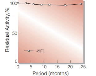
Fig.1. Stability (Powder form)
(kept under dry conditions)
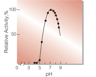
Fig.2. pH-Activity
37℃,15 min-reaction in BrittonRobinson buffer
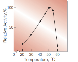
Fig.3. Temperature activity
15min-reaction in 0.1M phospate buffer, pH7.3
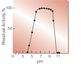
Fig.4. pH-Stability
25℃,20hr-treatment with BrittonRobinson buffer
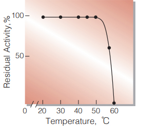
Fig.5. Thermal stability
15min-treatment with 50mM phosphate buffer,pH7.3 contg. 1.0mM MgCl2 enzyme concn.:80U/mL
| Size | 1 MG, 10 MG, 5 MG |
|---|
10 in stock
10 in stock
10 in stock
10 in stock


Reviews
There are no reviews yet.