-dependent) from Microorganism")
| Appearance | White amorphous powder, lyophilized. | |
|---|---|---|
| Activity | GradeⅢ 250 U/mg-solid or more | |
| Contaminants | NADH oxidase | ≤1.0×10⁻³ % |
| α-Glucosidase | ≤1.0×10⁻³ % | |
| Glucose-6-phosphate dehydrogenase | ≤1.0×10⁻³ % | |
| Stability | Stable at −20 ℃ for at least one year(Fig.1) | |
|---|---|---|
| Molecular weight | approx. 101,000 (Gel filtration) | |
| Isoelectric point | 4.5 | |
| Michaelis constants | NAD+linked | 1.38×10⁻² M (D-Glucose) 3.09×10⁻⁴ M (NAD+) |
| NADP+linked | 1.25×10⁻² M (D-Glucose) 4.07×10⁻⁵ M (NADP+) | |
| Inhibitors | Ag+, Hg2+, Monoiodoacetate | |
| Optimum pH | 9.0(Fig.4) | |
| Optimum temperature | 55 ℃(Fig.5) | |
| pH Stability | pH 6.0−7.5 (20 ℃, 16 hr)(Fig.6) | |
| Thermal stability | 45 ℃ (15 min-treatment with 50 mM K-phosphate buffer, pH 7.0)(Fig.7) | |
| Substrate specificity | Specific for β-D-Glucose or 2-Deoxy-glucose (Table.1) (Either NAD+ or NADP+ serves as coenzyme.) | |
This enzyme is useful for enzymatic determination of D-glucose.
![]()
The formation of NADH is measured at 340 nm by spectrophotometry.
One unit causes the formation of one micromole of NADH per minute under the conditions detailed below.
| A. Tris-HCl buffer, pH 8.0 | 0.1 M |
|---|---|
| B. D-Glucose solution | 1.5 M |
| C. β-NAD+ solution | 80 mg/mL |
| D. Enzyme diluent | 50 mM K-phosphate buffer, pH 7.0 contg. 0.1 % BSA |
1.Prepare the following reaction mixture in a cuvette (d = 1.0cm) and equilibrate at 37 ℃ for approximately 5 minutes.
| 2.6 mL | Tris-HCl buffer, pH 8.0 | (A) |
| 0.3 mL | Substrate solution | (B) |
| 0.1 mL | β-NAD+ solution | (C) |
| Concentration in assay mixture | |
|---|---|
| Tris-HCl buffer | 85.25 mM |
| D-Glucose | 147.54 mM |
| NAD+ | 3.66 mM |
2.Add 0.05 mL of the enzyme solution* and mix by gentle inversion.
3.Record the increase in optical density at 340 nm against water for 2 to 5 minutes with a spectrophotometer thermostated at 37 ℃, and calculate the ΔOD per minute from the initial linear portion of the curve (ΔOD test).
At the same time, measure the blank rate (ΔOD blank) using the same method as the test except that the enzyme diluent (D) is added instead of the enzyme solution.
*Dissolve the enzyme preparation in ice-cold enzyme diluent (D), dilute to 0.8−1.2 U/mL with the same buffer and store on ice.
Activity can be calculated by using the following formula :
Volume activity (U/mL) =
ΔOD/min (ΔOD test−ΔOD blank)×Vt×df
6.22×1.0×Vs
= ΔOD/min×9.807×df
Weight activity (U/mg) = (U/mL)×1/C
| Vt | : Total volume (3.05 mL) |
| Vs | : Sample volume (0.05 mL) |
| 6.22 | : Millimolar extinction coefficient of NADH under the assay conditions (cm2/micromole) |
| 1.0 | : Light path length (cm) |
| df | : Dilution factor |
| C | : Enzyme concentration in dissolution (c mg/mL) |
| Substrate (150mM) | Relative activity(%) |
|---|---|
| D-Glucose | 100.0 |
| L-Glucose | 0.0 |
| D-Xylose | 16.2 |
| 2-Deoxy-glucose | 127.0 |
| L-Sorbose | 0.0 |
| D-Mannose | 5.1 |
| D-Fructose | 0.0 |
| Substrate (150mM) | Relative activity(%) |
|---|---|
| Galactose | 1.7 |
| D-Lactose | 1.5 |
| D-Sorbitole | 0.0 |
| D-Mannitol | 0.0 |
| Sucrose | 0.0 |
| Inositol | 0.0 |
| Maltose | 1.4 |
[The enzyme dissolved in 50 mM K-phosphate buffer, pH 7.0 (2.8 U/mL) was incubated with each chemical for 1 hr at 30 ℃.]
| Chemical | Concn.(mM) | Residual activity(%) |
|---|---|---|
| None | – | 100 |
| Metal salt | 2.0 | |
| AgNO3 | 7.1 | |
| Ba(OAc)2 | 98.2 | |
| CaCl2 | 98.9 | |
| Cd(OAc)2 | 96.6 | |
| CoCl2 | 96.4 | |
| CuSO4 | 99.5 | |
| FeCl3 | 98.1 | |
| FeSO4 | 96.6 | |
| HgCl2 | 5.9 | |
| MgCl2 | 101.5 | |
| MnCl2 | 100.9 | |
| NiCl2 | 93.4 | |
| Pb(OAc)2 | 99.8 | |
| ZnSO4 | 102.1 |
| Chemical | Concn.(mM) | Residual activity(%) |
|---|---|---|
| KF | 2.0 | 98.7 |
| NaF | 10.0 | 100.6 |
| NaN3 | 20.0 | 101.6 |
| NEM | 2.0 | 97.6 |
| MIA | 2.0 | 0.4 |
| IAA | 2.0 | 92.2 |
| EDTA | 5.0 | 107.2 |
| (NH4)2SO4 | 20.0 | 96.0 |
| Borate | 20.0 | 101.4 |
| o-Phenanthroline | 2.0 | 97.7 |
| α,α′-Dipyridyl | 1.0 | 100.3 |
| Urea | 2.0 | 122.5 |
| Guanidine | 2.0 | 99.2 |
| Hydroxylamine | 2.0 | 107.2 |
Ac, CH3CO; NEM, N-Ethylmaleimide; MIA, Monoiodoacetate; IAA, lodoacetamide; EDTA, Ethylenediaminetetraacetate.
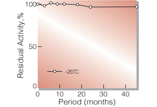
Fig.1. Stability (Powder form)
(kept under dry conditions)
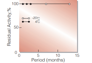
Fig.2. Stability (Powder form)
(kept under dry conditions)
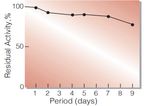
Fig.3. Stability (Liquid form)
25 ℃,in 83 mM Tris-HCI buffer solution pH 8.0(contg.3.7 mM β-NAD,40 U/mL mutarotase) enzyme concn.:300 U/mL
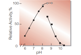
Fig.4. pH-Activity
37 ℃,5 min-reaction in 80 mM buffer solution
●:pH 6.0-8.0 K-phosphate
○:pH 8.0-9.0,Tris-HCI
■:pH 8.5-10.5 Carbonate
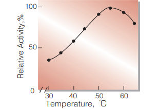
Fig.5. Temperature activity
(in 80 mM Tris-HCI buffer, pH 8.0)
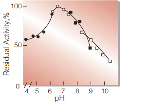
Fig.6. pH-Stability
20 ℃,16 hr with 0.1 M buffer solution
●:pH 4.0-6.0 acetate
○:pH 6.0-8.0 K-phosphate
■:pH 7.5-9.0 Tris-HCI
□:pH 8.5-10.5 carbonate
enzyme concn.:10 U/mL
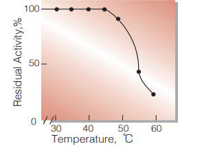
Fig.7. Thermal stability
15 min-treatment with 50 mM K-phosphate buffer pH 7.0 enzyme concn.: 12 U/mL
| Size | 1 MG, 10 MG, 5 MG |
|---|
10 in stock
10 in stock
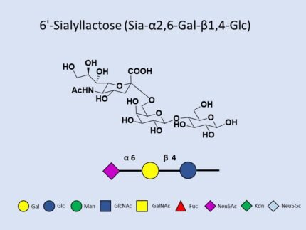
Catalog No: G01534TU
CAS No: 64309-01-9
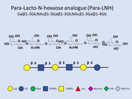
Molecular Formula: C₅₂H₈₈N₂O₃₉
Molecular Weight: 1365.25 g/mol
Packaging: Available in research quantities
10 in stock
10 in stock

Reviews
There are no reviews yet.